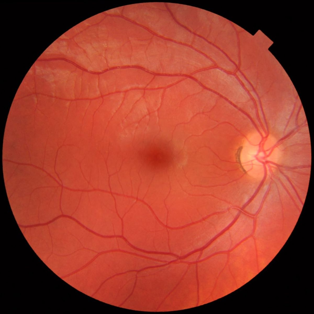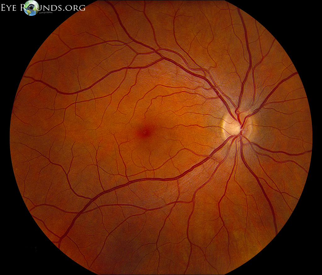

Examination of retina (fundus examination) is an important part of the general eye examination. dilating the pupil using special eye drops greatly enhances the view and permits an extensive examination of peripheral retina. Apr 17, 2021 · large aperture: used for viewing the fundus through a dilated pupil and for the general examination of the eye slit aperture : can be helpful in assessing contour abnormalities of the cornea, lens and retina as it makes elevation easier to see. Mar 16, 2021 reduced vision: examination should cover the whole visual/refractory axis from cornea to fundus, with functional testing of pupils, optic nerve . Feb 28, 2019 ophthalmoscopy is an examination of the back part of the eye (fundus), which includes the retina, optic disc, choroid, and blood vessels.
Jul 15, 2015 ocular surgery news a 26-year-old female nursing school graduate was referred to the new england eye center for decreased vision in both . The direct ophthalmoscope allows you to look into the back of the eye to look at the health of the retina, optic nerve, vasculature and vitreous humor. this exam . Fundal examination should be an integral part of any eye examination. the cup/ disk ratio is slightly larger in the african american population. the normal fundus . See more videos for fundus examination eye.

Look at right fundus with fundus examination eye your right eye ophthalmoscope should be close to your eyes. your head and the scope should move together set the lens opening at +8 to +10 diopters. Traditionally, most neurologists would have used a direct ophthalmoscope to examine the ocular fundus (optic nerve, macula, and retina and vessels of the posterior pole of the eye). however, ocular funduscopy appears to be a dying art,1 and emerging technologies, such as non-mydriatic ocular fundus cameras and smartphone attachments may make the direct ophthalmoscope obsolete. 2. Fundus american academy of ophthalmology the fundus is the inside, back surface of the eye. it is made up of the retina, macula, optic disc, fovea and blood vessels. the fundus is the inside, back surface of the eye.
Childhood Eye Examination American Family Physician
Sep 29, 2020 · fundus photography is the process of taking serial photographs of the interior fundus examination eye of your eye through the pupil. a fundus camera is a specialized low-power microscope attached to a camera used to examine structures such as the optic disc, retina, and lens. Of a clinician's coat and can be used in settings where other methods of ocular fundus examination are unavailable; (3) relatively low cost; (4) its practicality as a .
Dilated fundus examination an overview sciencedirect topics.
Clinical examination of the ocular fundusrichard f. spaide and silvana negrao many kinds of diseases can affect the posterior segment of the eyes. to make a correct diagnosis and establish an adequate treatment, one must combine a proper history of the patient’s symptoms with a detailed ocular examination.
Fundoscopic / ophthalmoscopic exam visualization of the retina can provide lots of information about a medical diagnosis. these diagnoses include high blood pressure, diabetes, increased pressure in the brain and infections like endocarditis. introduction to the fundoscopic / ophthalmoscopic exam. Ophthalmoscopy is a test that allows your ophthalmologist, or eye doctor, fundus examination eye to look at the back of your eye. this part of your eye is called the fundus, and consists . For example, immediately after the paragraph cited on the dilated fundus examination, one reads. the components of ocular health assessment and systemic . Funduscopic examination is a routine part of every doctor's examination of the eye, not just the ophthalmologist's. it consists exclusively of inspection. one looks through the ophthalmoscope (figure 117. 1), which is simply a light with various optical modifications, including lenses.
State of illinois eye examination report recommendations 1. correctivelenses: no yes,glassesorcontactsshouldbewornfor: constantwear nearvision farvision. Anatomy the part of a hollow organ that is farthest away from the organ's opening; the fundus examination eye bladder, gallbladder, stomach, uterus, eye, and middle ear cavity all have a fundus ophthalmology the interior of the eyeball–ie, retina, optic disc, macula, which is seen during a fundoscopic eye examination.

Apr 27, 2017 the examiner moves the lens slightly toward or away from the eye until the image of the fundus completely fills the lens. the magnification . Dilated fundus examination or dilated-pupil fundus examination (dfe) is a diagnostic procedure that employs the use of mydriatic eye drops (such as tropicamide) to dilate or enlarge the pupil in order to obtain a better view of the fundus of the eye.
Ophthalmoscopy Medlineplus Medical Encyclopedia
Dec 19, 2016 · ophthalmoscopy is a test that allows your ophthalmologist, or eye doctor, to look at the back of your eye. this part of your eye is called the fundus, and consists of: retina. Mar 16, 2021 · reduced vision: examination should cover the whole visual/refractory axis from cornea to fundus, with functional testing of pupils, optic nerve and macula. if visual loss is stated or detected or there are neurological symptoms, visual fields should always be checked. Visual acuity, confrontational visual fields, pupils, and ocular motility should be assessed in all patients with a history of trauma. if possible, slit lamp examination . Apr 17, 2021 large aperture: used for viewing the fundus through a dilated pupil and for the general examination of the eye; slit aperture: can be helpful in .
Funduscopic examination includes the optic nerve (specifically, checking for cup-to-disc ratio, edema, and pallor), the retina around the optic nerve, the macula (specifically, checking for color, edema, hemorrhages, exudates, and masses), and arteries and veins (specifically, checking for size, occlusion, and emboli) (fig. 2. 4). Look at right fundus with your right eye; ophthalmoscope should be close to your fundus examination eye eyes. your head and the scope should move together; set the lens opening at +8 to +10 diopters. with the ophthalmoscope 12-15 inches from the patient's eye, check for the red reflex and for opacities in lens or aqueous. Aug 15, 2013 · a symmetric orange-red light should reflect from each fundus; however, the tint may vary to light gray with darkly pigmented eyes. eye examination in infants, children, and young adults by.
0 Response to "Fundus Examination Eye"
Posting Komentar